Dermatofibroma
Section shows dermis which shows tumour tissue that is composed of proliferating fibroblasts present in interlacing bundles. Tumour also shows a historiform pattern.

Lichen Planus
Section show laminated hyperkeratosis with irregular acanthosis of the epidermis. The retepegs are elongated and pointed giving a saw toothed appearance to the section.Wedge shaped hypergranulosis is present and there is marked degeneration of the basal layer with presence of several civatte bodies at the dermo-epidermal junction.A band like infiltrate of lymphocytes and histiocytes is presene in the superficial dermis aborting to the epidermis.

Lichen Planus Hypertrophicus
Section shows laminated hyperkeratosis with patchy parakeratosis and marked keratotic plugging.The epidermis is irregularly acanthotic with elongated thickened , bulbous retepegs. There is wedge shaped hypergranulosis seen and patchy degeneration of the basal cell layer is present. A band like infiltrate of lymphocytes and histiocytes is seen in the superficial dermis aborting on to the epidermis at places.
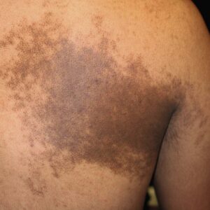
Becker Nevus
Section shows mild basket weave, hyperkeratosis, the epidermis is acanthotic with downward extension and branching of the rete pegs. The granular layer is present throughout and basal cell layer is intact with focal hyperpigmentation specially at the lower most end of the rete pegs. The dermis shows no pathology.
Intradermal Nevus
Section shows mild laminated hyperkeratosis with atrophy of the epidermis. The rete peg pattern is well maintained. Basal cell layer is intact with normal pigmentation. No functional activity seen. Dermis shows presence of tumor tissue composed of nests and cords of proliferating melanocytes. Nests of cells in the superficial dermis are round with central nuclei and abundant melanin pigment. The tumor cells in the deeper dermis are spindle shaped arranged in bundles simulating nerve fibres. No atypical changes seen.
Sweet Syndrome
Section shows mild basket weave hyperkeratosis with irregular acanthosis of the epidermis. The basal cell layer is intact with normal pigmentation. A sub-epidermal bulla is present which shows fibrin strands with few neutrophils. The base of the bulla is formed by the dermis that shows a band like infiltrate of neutrophils with few lymphocytes and histiocytes. There is marked superficial edema with dialatation and congestion of blood vessels.
Schamberg’s disease
Section shows laminated hyperkeratosis with karatotic plugging. The epidermis is atrophic and shows a complete flattening of the rete pegs. The granular layer is present throughout and the basal cell layer is intact with normal pigmentation. A band like infiltrate of lymphocytes and histiocytes is present in the superficial dermis.
Perl’s stain is positive for iron deposition.
Milia
Section shows laminated hyperkeratosis overlying an epidermis that shows patchy atrophy. There are several cystic structures present that are arising from the lower portion of the infundibulum of the vellus hairs. The cysts are lined by stratified epithelium and contain concentric ring of keratin. A sparse perivascular lymphocytes is present in the surrounding blood vessels.
Basal Cell Carcinoma
Section shows mild basket weave pattern of stratum corneum with follicular plugging . the epidermis is atrophic with flattening of rete pegs. The dermis shows presence of tumor tissues present in lobules that are surrounded by proliferating fibroblasts. Tumor cells have a monomorphic appearance and shows large hyperchromatic nuclei surrounded by thin rim of cytoplasm. Palisading mass present in the groups of tumor cells among which some shows central keratinisation.
Hansen’s BL
Section shows laminated hyperkeratosis. The epidermis is mildly acanthotic with short and broad rete ridges. Granular layers are present throughout and basal layer is intact with normal pigmentation. Dermis shows a no. of epithelioid cells and histiocytes with a few Langhan’s giant cells. A no. of foamy macrophages can also be seen in the same area. FF stain is negative for AFB.
LD Bodies
BT
Section shows granulomas with peripheral lymphocytes that follow the neuro-vascular bundles and infiltrate the sweat glands and erector pilli muscles. Langhan’s giant cells are variable in no.
Morphea
Section shows mild basket weave hyperkeratosis. The epidermis is normal in appearance and has a well defined rete peg pattern. Basal cell layer is intact and shows normal pigmentation. There is thickening and hyalinization of the dermal collagen, together with a perivascular lympho-hystiocytic infiltrate seen in the superficial dermis. The sweat glands are atrophic and seem to be “pulled up: into the dermis.
VVG stain shows normal elastic fibers.
Fiteferrako stain showing numerous acidfast bacilli in clumps
Spongiosis
Epidermis showing spongiosis and spongiotic vesicle.
Pigmented Seborrhoeic Keratosis
Section shows a well defined lesion consisting of hyperkeratosis ,acanthosis and papillamatosis of the epidermis. Irregular download proliferation of the rete pegs are seen enclosing a number of horn cysts filled with keratin. The basal cell layer is intact with focal hyperpigmentation. A sparse perivascular lymphohistiocytic infiltrate is seen in superficial dermis.
Epidermis showing many basiloid cell proliferation.
Pityriasis lichenoides
Lichen planus
Section shows laminated hyperkeratosis with irregular acanthosis of the epidermis. Retepegs are elongated and pointed giving a saw toothed appearance to the section. Wedge shaped granulosis is present and there is extensive basal cell degeneration seen. A band like infiltrate of lymphocytes and histiocytes is present in the superficial dermis closely abutting onto the epidermis.
Histoid leprosy
Section shows laminated hyperkeratosis overlying a mildly atrophic epidermis. There is flattening of rete pegs but the basal layer is intact with normal pigmentation. A free grenz zone is present beneath which the dermis shows infilteration by foamy histiocytes and few lymphocytes. The histiocytes at places show spindled out appearance and there is evidence of nerve infilteration. FF stain is positive for AFB.
Sarcoidosis
Section shows laminated hyperkeratosis with mild atrophy of the epidermis. There is partial flattening of the rete pegs but the basal cell layer is intact and shows normal pigmentation. Several well defined granulomas of epitheloid and giant cells are present in the dermis. Sprinkling of lymphocytes seen in between granulomas though majority of them appear to be naked. Occasional granulomas shows central areas of necrosis. No evidence of nerve infiltration seen.
Lichen sclerosus et atrophicus
Section shows laminated hyperkeratosis overlying an atrophic dermis. There is complete flattening of the rete pegs and marked basal cell degeneration with sub-epidermal clefting at places. The superficial dermis is oedematous and has a “groundglass” appearance.a perivascular lymphohistiocyte infiltrate is seen in mid dermis.
Syringoma
Section shows basket weave pattern of stratum corneum overlying a normal epidermis with well defined rete peg pattern. Basal layer is intact and shows normal pigmentation. The dermis shows the presence of tumor tissue that is made up of several tubular structures embedded in a fibrous stroma. These ducts are lined by two rows of epithelial cells with the inner layer showing marked vacuolization. Amorphous material is present in some of these ducts that also posses a small comma like.
Lupus vulgaris
Section shows laminated hyperkeratosis with follicular plugging. The epidermis is mildly atrophic and shows flattening of the rete pegs. Focal vacuolar degeneration of the basal layer is present. A granulamatous infiltrate is seen in the dermis. It is made of collection of epitheloid cells with occasional langerhans giant cells that are surrounded by large number of lymphocytes and few plasma cells.
Tricholemmal cyst
Section shows basket weave hyperkeratosis with mild acanthosis with elongated rete pegs. The dermis shows a no. of cysts lined by stratified squamous epithelium, containing a no. of sebaceous glands and a few follicular infundibula. The cyst wall shows abrupt tricholemmal keratinization. The cyst wall is rich in glycogen and cysts contain many cholesterol clefts
Squamous cell carcinoma
Section shows hyperkeratotic acanthotic stratified squamous epithelium. The underlying subepithelial tissue shows the presence of groups and nests of malignant squamous cells infilterating up to and into the muscle layers. The cells are large with central hyperchromatic nuclei and there are areas of keratinization seen with formation of squamous perls. Few mitotic figures are present and the surrounding connective tissue is heavily infilterated by lymphocytes, histiocytes, eosinophils and few neutrophils.
DH
Section shows basket weave pattern of stratum corneum and mild acanthosis of epidermis. There is large sub-epidermal bulla that is filled with fibrin strands in which are present mainly neutrophils and few eosinophils. The dermal papillae adjacent to bullae shows marked papillary oedema and formation of microabscesses that are heavily infiltrated with neutrophils. A perivascular lymphohistiocytic infiltrate with neutrophils and eosinophils are present in epidermis.
Postular psoriasis
Lymphangioma
Psoriasis
Section shows hyperparakeratosis with general aggregates of neutrophils forming numerous abscesses. The epidermis is acanthotic and has narrow elongated rete pegs. Marked hypogranulosis is seen but basal cell layer is intact and shows normal pigmentation. There is papillary edema and thinning of the suprapapillary epidermal plates. Thin walled congested blood vessels are present in dermal papillae and an perivascular lymphohistiocyic infilteration is present in superficial dermis.
Bullous pemphigoid
Pityriasis lichenoides chronicus
Section shows hyperparakeratosis with mild acanthosis of the epidermis. The retepegs are elongated and narrow. There is extensive hypogranulosis but basal cell layer is intact and shows normal pigmentation. Dermal papillae shows several thin walled congested vessels and a sparse perivascular lymphohistiocytic infiltrate is present in superficial dermis.
Discoid lupus erythematosus
Section shows laminated hyperkeratosis with marked keratotic and follicular plugging. The epidermis is atrophic with flattening of rete pegs at places. There is patchy vacuolar degeneration of the basal layer with thickening and tortuosity of the basal membrane. Perivascular and perappendageal lymphocytic infiltrate are present throughout the dermis. Focal mucinous degeneration of dermal collagen is also seen.
Keloid

 Dr Sachin k Maurya, Medical Director, Antiaging skin clinic Group